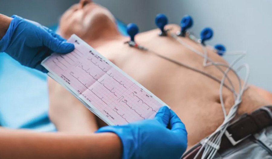
ECG (Electrocardiography)

Electrocardiography (ECG or EKG) is a crucial diagnostic tool in cardiology that provides detailed information about the heart’s electrical system. The heart’s rhythm, rate, and overall function are governed by electrical impulses. These impulses travel through the heart muscle and coordinate its contractions, allowing blood to flow efficiently throughout the body. An ECG records these electrical impulses, helping healthcare professionals diagnose various heart-related conditions.
How ECG Works
An ECG measures the electrical activity of the heart by placing electrodes on the skin. These electrodes detect the electrical signals generated by the heart as it beats and transmit them to the ECG machine. The signals are then displayed as a series of waves on a graph. These waves represent different phases of the heart’s electrical cycle:
- P Wave: Represents the electrical impulse as it travels through the atria, leading to atrial contraction.
- QRS Complex: Represents the electrical impulse as it travels through the ventricles, causing ventricular contraction.
- T Wave: Represents the recovery phase, when the ventricles relax and the heart prepares for the next beat.
Types of ECG
There are different types of ECG tests, each suited for specific diagnostic purposes:
- Resting ECG: Performed while the patient is lying down and relaxed. This is the most common type and helps assess the heart’s basic electrical activity.
- Stress ECG (Exercise ECG): Performed while the patient exercises on a treadmill or stationary bike. This test helps assess the heart’s function under physical stress and is used to diagnose conditions like coronary artery disease.
- Holter Monitoring (Ambulatory ECG): This is a 24- to 48-hour ECG recording using a portable device that the patient wears. It provides a more detailed analysis of the heart’s activity over an extended period, helping diagnose intermittent arrhythmias that may not appear during a standard ECG.
- Event Monitor: Similar to Holter monitoring but used for longer periods (up to several weeks). It records the heart’s activity at times the patient triggers manually when they experience symptoms like chest pain or palpitations.
- Telemetry ECG: A wireless ECG that continuously monitors the heart’s electrical activity, typically used in hospital settings, especially for critically ill patients or those undergoing heart surgery.
Indications for an ECG
An ECG is often recommended for individuals who present with certain symptoms or risk factors. Some common reasons for an ECG include:
- Chest pain or discomfort
- Shortness of breath
- Palpitations (irregular heartbeats)
- Dizziness or fainting
- Fatigue or weakness
- High blood pressure
- History of heart disease or family history of cardiac conditions
- Monitoring for heart disease progression in known patients
Common Heart Conditions Diagnosed Using ECG
- Arrhythmias: Irregular heart rhythms, such as atrial fibrillation, ventricular tachycardia, or bradycardia (slow heart rate), can be identified through abnormal ECG patterns.
- Heart Attacks (Myocardial Infarction): An ECG can show the characteristic changes in electrical activity caused by a heart attack, such as ST-segment elevation or abnormal T waves.
- Cardiac Ischemia: Reduced blood flow to the heart muscle can cause changes in the ECG, often leading to a diagnosis of ischemia.
- Heart Enlargement (Cardiomegaly): An ECG can help detect changes in the heart’s electrical activity that occur as a result of enlarged heart chambers.
- Electrolyte Imbalances: Abnormal levels of potassium, calcium, or sodium can affect the heart’s electrical activity, often detectable on an ECG.
- Conduction Disorders: ECG is used to assess conditions such as heart block, where the electrical impulses are delayed or blocked as they pass through the heart.
- Pericarditis: Inflammation of the heart’s lining can produce characteristic changes in the ECG.
Advantages of ECG
- Non-invasive and painless: No needles or injections are involved, and the procedure is quick, often taking only 5 to 10 minutes.
- No radiation: Unlike some imaging tests, an ECG does not use radiation.
- Real-time monitoring: Immediate results help healthcare providers assess the heart’s condition in real time.
- Cost-effective: ECGs are relatively inexpensive compared to other diagnostic imaging tests.
- Widely available: ECG machines are found in most hospitals, clinics, and even many general practitioners’ offices.
Limitations of ECG
While an ECG is an essential diagnostic tool, it does have some limitations:
- Limited information: It provides only electrical data and does not offer detailed structural information about the heart. For this, imaging tests like echocardiography or MRI are needed.
- Not always conclusive: Some heart conditions may not show up on an ECG, particularly if the problem is intermittent or occurs during certain activities.
- Requires interpretation: A trained healthcare professional is necessary to interpret the ECG results accurately, as abnormalities can sometimes be subtle or similar to other conditions.
